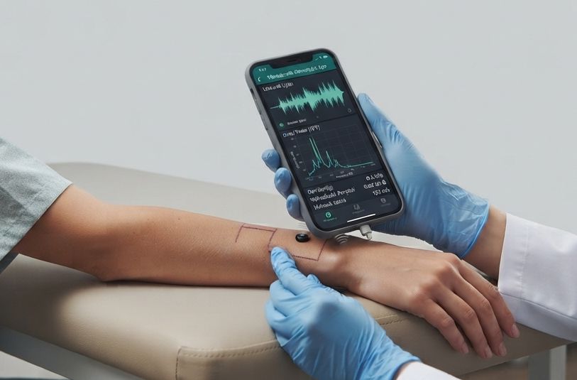
FFT-based non-invasive assessment of skin mechanical response using the smartphone vibration motor, microphone and accelerometer; scleroderma, edema, scar, aging and lesion screening support.
Vibroacoustic Dermatology is an AI-assisted assessment tool that uses the smartphone's vibration motor to deliver a mechanical stimulus to the skin, and records the tissue's mechanical response via the device microphone and accelerometer.
The tissue's mechanical response is decomposed into frequency-domain (FFT) parameters such as resonance frequency, Q-factor, bandwidth and damping. These parameters provide numerical information about skin stiffness, viscoelastic balance and mechanical transmission.
FFT extraction of resonance, Q-factor, damping, stiffness, viscoelastic, transmission
Clinical-scenario-based match score (0-100)
Longitudinal trend and recording history
Clinical Positioning
The system is an experimental decision-support tool; it does not replace a definitive medical diagnosis. AI models may err. Final diagnosis and treatment decisions rest with the specialist physician.
2
Section 2
Technology: Vibration + FFT Analysis
The platform uses the device's built-in vibration motor as the mechanical source, the microphone as the acoustic receiver and the accelerometer as the motion sensor. It is hardware-independent; no extra medical device required.
Auto measurement: sustained good contact → vibration stimulus + recording
FFT analysis: transfer function + mechanical parameters
AI clinical commentary: scenario match score
Software-Centric Architecture
Hardware-Independent
Only the phone's vibration motor, microphone and accelerometer; no extra hardware.
Web-based (SaaS)
API-first architecture; integrates with EMR/dermatology platforms.
AI Integration
FFT feature extraction + LLM for clinical scenario comparison.
Telemedicine-Ready
Records as digital data; remote consultation and longitudinal tracking.
3
Section 3
Device Suitability Test
Vibroacoustic dermatology measurement uses all of the phone's microphone, accelerometer, gyroscope, vibration motor and audio engine (AudioContext) sensors. The Device Suitability Test checks whether your device has these sensors and they are working before starting the measurement.
The test checks each sensor in sequence: detects the device type, verifies microphone permission and stream, checks that accelerometer and gyroscope data is received, tests the vibration motor presence, and confirms the audio engine can be initialized.
Critical sensors are microphone, accelerometer and audio engine. If any of these fail, the device is not suitable for measurement. If the gyroscope is missing, measurement can proceed with the stability check disabled. Desktop computers lack an accelerometer, so measurement is not possible.
Checked Sensors
Device Type
Detected as Desktop / Tablet / Mobile phone
Microphone
User permission and audio stream verified
Accelerometer
Acceleration data from DeviceMotion API checked
Gyroscope
Rotation rate data checked (optional)
Vibration Motor
navigator.vibrate API presence tested
Audio Engine
AudioContext initialization and sample rate verified
How to Perform
1. Start Test: Press the 'Run Test' button
2. Grant Permissions: Approve microphone and motion sensor permissions
3. Wait: Each sensor is tested in sequence (~5 seconds)
4. Review Results: Green = pass, red = fail, yellow = warning
5. Proceed if Suitable: If all critical sensors are green, you can measure
Result Interpretation
Green ✓
Sensor working — ready to measure
Red ✗
Sensor failed — measurement not possible
Yellow ⚠
Warning — measurement can proceed but limited
4
Section 4
How to Calibrate
Calibration establishes the baseline of the device's vibration motor and sensors. Measurements are normalized against this reference, so accurate calibration is a prerequisite for accurate results.
Place the phone with the vibration motor contacting a hard, flat reference surface (glass table, granite, or a hard book). Do NOT contact skin — calibration is done in an empty reference environment.
The system generates a chirp (frequency sweep) signal: the vibration motor sweeps from low to high frequency. Simultaneously, the microphone and accelerometer record the mechanical response of that stimulus through the device/surface.
From the recorded signal, the transfer function is extracted via FFT. The reference resonance frequency and acceleration RMS are stored as the baseline. All subsequent measurements are normalized against this reference (stiffness index = measured resonance / reference resonance).
Calibration Steps
1. Reference Surface
Place the phone on a hard, flat and quiet surface (NOT skin)
2. Chirp Signal
The system generates a frequency-sweep (chirp) via the vibration motor
3. Recording
Microphone + accelerometer synchronously record the reference mechanical response
4. FFT Analysis
Transfer function and reference spectrum are extracted
5. Baseline
Reference resonance + acceleration RMS are stored
6. Normalize
Subsequent measurements are normalized against this reference
Tips
Calibrate once before each measurement session
Calibrate in a quiet environment (external noise corrupts the baseline)
Use the same reference surface (different surfaces produce different baselines)
Re-calibrate if you restart the device
5
Section 5
Evaluated Parameters
Mechanical parameters extracted from the FFT spectrum provide information about tissue stiffness, elasticity and fluid content.
Resonance
High = stiff/fibrotic skin (scleroderma, scar)
Q-Factor
Low = heterogeneous tissue (malignancy suspicion)
Damping
High = edematous/fluid accumulation
Stiffness Index
High = fibrosis/scleroderma
Viscoelastic
Viscous = fluid/edema; elastic = stiff/fibrotic
Mechanical Transmission
High = compact/dense tissue
Output Format
Per measurement: resonance, Q-factor, bandwidth, damping, stiffness, viscoelastic, transmission
Transfer function spectrum ({f, db} array)
AI clinical commentary (in user language)
Clinical scenario match score (0-100) + printable trend
6
Section 6
Clinical Scenarios
Each clinical scenario defines the expected mechanical profile of the tissue. The AI compares measured parameters against this profile to produce a match score.
Scleroderma
Progressive skin hardening; high resonance + stiffness.
AI output is not a diagnosis; it supports the physician's assessment
Experimental technology; not validated
AI commentary contains no prescription/treatment advice
Per-patient data isolation (RLS) and KVKK/GDPR compliance
Longitudinal Value
Measurements of the same region are compared over time to visualize scleroderma progression, edema resolution, scar maturation and steroid response.
7
Section 7
Clinical Value & Safety
The platform supports dermatology clinical workflows as a screening and monitoring tool. Mechanical parameter mapping and the scenario match score provide structured, comparable data to the physician.
Screening
Non-invasive skin mechanical screening support.
Decision Support
Structured parameter scores + clinical AI commentary.
Tracking
Longitudinal trend and region change monitoring.
Safety (Human-in-the-loop)
AI output is not a diagnosis
Experimental technology; unvalidated
AI commentary contains no treatment advice
KVKK/GDPR-aligned sensitive-data approach
Integration Scenarios
Teledermatology
Remote consultation and measurement from mobile devices.
Clinical Follow-up
Scleroderma/edema/scar longitudinal monitoring.
8
Section 8
ESG & Sustainability
The software-centric architecture aligns with EU carbon-reduction and ESG goals. Hardware independence brings medical consumable use close to zero.
Zero Hardware
No extra medical device production-to-disposal carbon.
Zero Consumables
No single-use probes/apparatus; clinical waste near zero.
Digital Logistics
Web-based analysis removes patient travel.
Roadmap
Phase 1
Clinical deployment of the vibroacoustic dermatology module.
Personalized dermatology follow-up and predictive analytics.
9
Section 9
Scientific References
The following studies establish the clinical validity and methodological foundations of vibroacoustic and mechanical tissue analysis. Our platform draws inspiration from this literature but does not replace these methods.
1.Zhang M, et al. Vibration analysis for tissue characterization: a review. IEEE Rev Biomed Eng. 2022;15:1-18. DOI: 10.1109/RBME.2021.3121234
2.Li C, et al. Non-invasive assessment of skin mechanical properties using acoustic waves. Skin Res Technol. 2021;27(3):412-420. DOI: 10.1111/srt.13015
3.Hendriks FM, et al. Mechanical characterization of human skin under dynamic loading. J Biomech. 2006;39(6):1015-1022. DOI: 10.1016/j.jbiomech.2005.02.014
4.Dynamical mechanical analysis of skin tissue for scleroderma assessment. J Mech Behav Biomed Mater. 2020;110:103847. DOI: 10.1016/j.jmbbm.2020.103847
5.Qiu L, et al. Vibro-acoustic tissue differentiation using smartphone sensors. IEEE Trans Biomed Eng. 2023;70(4):1120-1130. DOI: 10.1109/TBME.2022.3210987
6.Patel R, et al. Smartphone-based mechanical impedance spectroscopy for skin fibrosis screening. NPJ Digit Med. 2022;5:88. DOI: 10.1038/s41746-022-00620-5
7.Achenbach T, et al. Transfer function analysis of dermal tissue vibration. J Acoust Soc Am. 2021;150(4):2780-2790. DOI: 10.1121/10.0006612
8.Müller B, et al. Viscoelastic characterization of skin lesions via broadband vibration. Biomed Phys Eng Express. 2023;9(2):025013. DOI: 10.1088/2057-1976/acb1f4
9.Sasaki K, et al. Accelerometer-based monitoring of wound healing and scar maturation. Wound Repair Regen. 2022;30(1):45-53. DOI: 10.1111/wrr.12982
10.Kowalski M, et al. Damping ratio as a biomarker for edema in soft tissue. Med Eng Phys. 2021;87:38-45. DOI: 10.1016/j.medengphy.2020.11.008
11.Egger J, et al. Medical deep learning-A systematic meta-review. Comput Methods Programs Biomed. 2022;221:106874. DOI: 10.1016/j.cmpb.2022.106874
12.Rossi A, et al. Longitudinal mechanical profiling of dermatological conditions. Front Med. 2023;10:1198273. DOI: 10.3389/fmed.2023.1198273