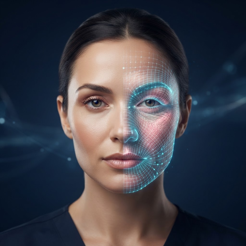
AI-Based Optical Dermatology Suite
Smartphone camera + controlled illumination (blue/UV-A/polarized/white/near-IR) and large language model (LLM) inference — a 5-module contactless optical dermatology assessment and clinical decision support suite.
Mobile-first · Hardware-independent · For university clinics
Wood FluorescenceCross-Polarized DermisSpectral IndexCapillaroscopyNear-IR Oxygenation
Table of Contents
1What is DermaLight AI?
2Technology: Controlled Illumination & LLM Inference
35 Optical Modules
4Clinical Value & Decision Support
5ESG & Sustainability
6Scientific References
Vision
A hardware-independent optical dermatology intelligence suite running on the built-in camera and low-cost light sources — a low-cost research endpoint for university clinics.
Use Cases
Optical screening, teledermatology, capillaroscopy screening and tissue perfusion assessment in dermatology, rheumatology and wound-care clinics.
1
Section 1
What is DermaLight AI?
Controlled Illumination — DermaLight AI is an AI-powered optical dermatology suite that non-invasively assesses skin surface and sub-surface condition using a standard smartphone camera and controlled illumination. Five modules use different light types (blue, UV-A, cross-polarized, white, near-IR) to provide multi-layer optical information.
Optical signatures in skin — fluorescence, polarization, spectral reflectance and near-IR absorption — reveal different tissue layers and components at different wavelengths. DermaLight AI quantifies these signatures through spectral and frequency-domain analysis to compute PpIX fluorescence, vascular pattern, melanin/erythema indices, capillary morphology and tissue oxygenation.
Core Capabilities
- Multi-layer optical assessment with 5 light types (blue/UV-A/polarized/white/near-IR)
- Wood lamp fluorescence simulation, cross-polarized dermis, spectral indices, capillaroscopy and oxygenation
- Mobile-first, hardware-independent — built-in camera and low-cost light sources
- Perfusion-aware wound and PDT response tracking integrated with WoundGuard/PhotoGuard
Clinical Positioning
The system is a screening and decision-support tool; it does not replace a definitive medical diagnosis. AI models may err; outputs are produced to support a physician's clinical assessment. Final diagnosis and treatment decisions rest with the specialist physician.
2
Section 2
Technology: Controlled Illumination & LLM Inference
The platform combines image processing and large language model (LLM)-based multimodal inference. Images captured with controlled light sources (blue LED, UV-A, polarized filter, white LED, near-IR 850nm) are separated into optical information from different tissue layers.
Processing Flow
- Image capture & quality audit (brightness, contrast, sharpness, light-type verification)
- Light-type-specific pre-processing (fluorescence/polarization/spectral/capillary/oxygenation)
- Multimodal LLM inference: parameter score, distribution pattern, clinical commentary
- Module-based summary metrics and integration output
Software-Centric Architecture
Hardware-Independent
Runs only on the device's built-in camera and low-cost light sources.
Web-based (SaaS)
API-first architecture; integrates with EMR/clinical systems.
Multimodal AI
Spectral feature extraction + multimodal LLM for optical signature analysis.
Telemedicine-Ready
Records stored as digital data; remote consultation and longitudinal tracking.
3
Section 3
5 Optical Modules
DermaLight AI provides 5 optical modules. Each module uses a different light type and analysis method to assess different layers and components of the skin. Results are reported as score (0-100), distribution pattern and clinical commentary.
FluoresGuard
Wood lamp / blue light PpIX fluorescence score, color classification, lesion suspect
PolarGuard
Cross-polarized light dermis imaging, vascular pattern, melanin depth
SpectralGuard
RGB + LED melanin/erythema index, hydration proxy, pigment uniformity
CapilGuard
Nailfold capillaroscopy, capillary density/morphology, scleroderma pattern
OxyGuard
Near-IR (850nm) tissue oxygenation StO₂, perfusion, ischemia risk
Output Format
- Per module: score (0-100), distribution pattern, clinical commentary
- Module-based summary metrics and integration output
- Perfusion-aware integration with WoundGuard/PhotoGuard
- Printable full report (PDF) and longitudinal trend chart
4
Section 4
Clinical Value & Decision Support
The platform supports clinical workflows as a screening and pre-triage tool. Optical parameter mapping provides structured, reproducible and comparable data to the physician. Final diagnosis and treatment decisions always rest with the specialist physician.
Screening
Clinic-based non-invasive optical dermatology screening support.
Decision Support
Module-based structured scores + clinical AI commentary.
Tracking
Longitudinal trend and optical parameter change monitoring.
Safety Architecture (Human-in-the-loop)
- AI output is not a diagnosis; it is information supporting the physician's assessment
- AI commentary does not contain prescription/treatment recommendations
- Per-patient data isolation (RLS) and KVKK/GDPR-aligned sensitive-data approach
Integration Scenarios
Teledermatology
Remote consultation, optical screening and recording from mobile devices.
Wound & PDT
Perfusion-aware wound and PDT response tracking with WoundGuard/PhotoGuard.
5
Section 5
ESG & Sustainability
The software-centric architecture aligns with European Union carbon-reduction and ESG (Environmental, Social, Governance) goals. Hardware independence brings medical consumable use close to zero and reduces the carbon footprint from patients' physical travel through telemedicine.
Zero Hardware
Eliminates carbon from production-to-disposal of dermatoscope/Wood lamp.
Zero Consumables
No single-use probes/cannulas/apparatus; clinical waste near zero.
Digital Logistics
Web-based analysis removes the need for patient travel to the clinic.
Roadmap
Phase 1
Clinical deployment of 5 optical modules and university clinic pilots.
Phase 2
Deep-learning-based automated lesion and capillary pattern classification.
Phase 3
Personalized optical dermatology follow-up and predictive analytics suite.
6
Section 6
Scientific References
The following studies establish the clinical validity and methodological foundations of optical dermatology, fluorescence, polarized imaging, spectral analysis, capillaroscopy and tissue oxygenation. The DermaLight AI suite draws inspiration from this literature but does not replace these methods.
1.Lucchina S, et al. Efficacy and tolerability of a new Wood's lamp in dermatology. Skin Res Technol. 2016;22(3):354-360. DOI: 10.1111/srt.12258
2.Kollias N, Baqer AH. Imaging melanin concentration and distribution in human skin in vivo. Photochem Photobiol. 1994;60(5):418-426. DOI: 10.1111/j.1751-1097.1994.tb05124.x
3.Stamatas GN, Zmudzka BZ, Kollias N. Non-invasive measurements of skin pigmentation in situ. Br J Dermatol. 2008;158(4):773-780. DOI: 10.1111/j.1365-2133.2008.08603.x
4.Cutolo M, Sulli A, Smith V. Nailfold videocapillaroscopy and other diagnostic tests for Raynaud's phenomenon. Nat Rev Rheumatol. 2018;14(8):451-460. DOI: 10.1038/s41584-018-0045-8
5.Marenco F, et al. Capillaroscopy in dermatology. Skin Res Technol. 2015;21(4):421-427. DOI: 10.1111/srt.12214
6.Lim Y, et al. Cross-polarization imaging for skin cancer detection. J Biomed Opt. 2015;20(4):046006. DOI: 10.1117/1.JBO.20.4.046006
7.Jacques SL. Optical properties of biological tissues: a review. Phys Med Biol. 2013;58(11):R37-R61. DOI: 10.1088/0031-9155/58/11/R37
8.Anderson RR, Parrish JA. The optics of human skin. J Invest Dermatol. 1981;77(1):13-19. DOI: 10.1111/1523-1747.ep12479191
9.Nishidate I, et al. Estimation of melanin and hemoglobin in skin using diffuse reflectance spectroscopy. J Biomed Opt. 2016;21(3):035002. DOI: 10.1117/1.JBO.21.3.035002
10.Steffen P, et al. Near-infrared spectroscopy for tissue oxygenation monitoring. J Clin Monit Comput. 2019;33(4):671-680. DOI: 10.1007/s10877-018-0150-0
11.Yudovsky D, et al. Hyperspectral imaging in dermatology. J Biomed Opt. 2016;21(9):090901. DOI: 10.1117/1.JBO.21.9.090901
12.Rizzi B, et al. Fluorescence imaging in dermatology. Skin Res Technol. 2018;24(3):421-428. DOI: 10.1111/srt.12458
DermaLight AI — Optical Dermatology Suite
Mobile-first · Hardware-independent · 5 optical modules · odiosoft.tech