NevusTrack — Brochure
NevusTrack
AI-Based Lesion Tracking Platform

NevusTrack - Lesion Tracking Platform

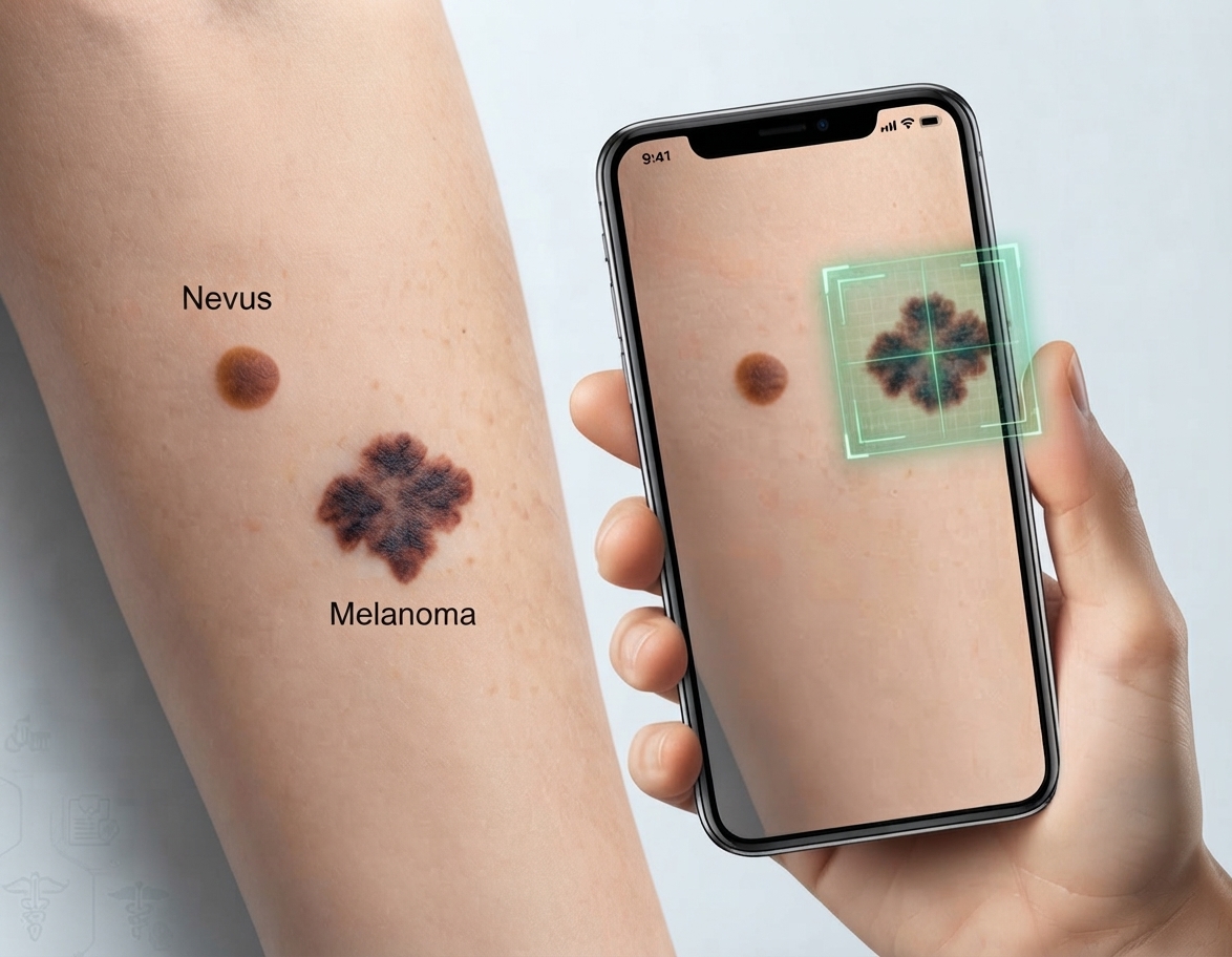
Contactless lesion photography with smartphone camera, AI-based ABCDE analysis, longitudinal tracking and differential diagnosis support.

Developed by Dr. Siyra Seren.
ABCDE AnalysisLongitudinal TrackingUV CorrelationHuman-in-the-loop
Table of Contents
1What is NevusTrack?
2ABCDE Criteria & AI Analysis
3Longitudinal Tracking & UV Correlation
4Clinical Value & Decision Support
5Safety & Sustainability
6Scientific References
Vision
A hardware-independent dermatology intelligence platform that produces clinically meaningful lesion assessment from a smartphone camera.
Use Cases
In-clinic screening, teledermatology, postoperative/medical skin follow-up and community-based early skin-cancer screening support.
1
Section 1

What is NevusTrack?

Lesion Tracking Platform — NevusTrack is a longitudinal monitoring system that uses AI to evaluate ABCDE criteria (Asymmetry, Border, Color, Diameter, Evolution) from lesion and nevus (mole) photographs. It works contactlessly with a standard smartphone camera; no extra medical device required.

The system evaluates ABCDE criteria such as diameter, pigment intensity, border irregularity and asymmetry from the lesion photo via AI. Changes of the same lesion region in the same patient over time can be compared; ∆ diameter, ∆ color and ∆ border metrics are computed automatically.

Core Capabilities

  • Contactless lesion photography with smartphone camera — no extra hardware
  • AI-based ABCDE analysis: Asymmetry, Border, Color, Diameter, Evolution
  • Longitudinal tracking: ∆ diameter, ∆ color, ∆ border metrics
  • Differential diagnosis panel: primary suspect, alternatives, confidence score
  • UV correlation: patient region / UV index risk stratification
Clinical Positioning
The system is a screening and decision-support tool; it does not replace a definitive medical diagnosis. AI models may err; outputs are produced to support a physician's clinical assessment. Final diagnosis and treatment decisions rest with the specialist physician.
2
Section 2

ABCDE Criteria & AI Analysis

The AI evaluates five ABCDE criteria from the lesion photo. Each criterion is reported with a 0-100 score, description and severity. A risk tier (low/moderate/high) and differential diagnosis panel are automatically generated.

ABCDE Criteria

Asymmetry (A)
Symmetry of the lesion halves — high asymmetry = suspicious
Border (B)
Border irregularity, notching — irregular = suspicious
Color (C)
Color variability / pigment intensity — multicolored = suspicious
Diameter (D)
Diameter (mm) and growth — >6mm or growing = suspicious
Evolution (E)
Change over time (longitudinal) — changing = suspicious

Processing Flow

  • Lesion photo capture & quality audit
  • AI ABCDE analysis: asymmetry, border, color, diameter, evolution
  • Risk tier: low / moderate / high
  • Longitudinal tracking: ∆ metrics and trend chart
  • Differential diagnosis panel + clinical AI commentary

Software-Centric Architecture

Hardware-Independent
Runs only on the phone camera; no dermatoscopy/probe required.
Web-based (SaaS)
API-first architecture; integrates with EMR/sleep platforms.
AI Integration
Image processing + multimodal LLM for ABCDE score and risk tier.
Telemedicine-Ready
Records stored digitally; remote consultation and longitudinal tracking.
3
Section 3

Longitudinal Tracking & UV Correlation

NevusTrack compares photographs of the same lesion over time to visualize growth, color and border changes. Trend charts and ∆ metrics are provided for each ABCDE criterion. An early warning is generated when flare or abnormal change is detected.

UV Correlation

A correlation analysis between the patient's geographic location and seasonal UV index versus lesion risk is performed. High UV exposure is assessed as a factor increasing melanoma risk and contributes to risk stratification.

Longitudinal Value
Photographs of the same lesion are compared over time to visualize growth/color/border changes, helping early detection of suspicious changes and reducing unnecessary biopsies.
4
Section 4

Clinical Value & Decision Support

The platform supports clinical workflows as a screening and pre-triage tool. ABCDE score, risk tier and differential diagnosis panel provide structured, reproducible and comparable data to the physician. Final diagnosis and treatment decisions always rest with the specialist physician.

Screening
Community/clinic-based non-invasive lesion screening support.
Decision Support
ABCDE score + risk tier + differential diagnosis panel.
Tracking
Longitudinal trend and lesion change monitoring.

Safety Architecture (Human-in-the-loop)

  • AI output is not a diagnosis; it is information supporting the physician's assessment
  • AI commentary does not contain prescription/treatment recommendations
  • Per-patient data isolation (RLS) and KVKK/GDPR-aligned sensitive-data approach

Integration Scenarios

Teledermatology
Remote consultation, postoperative skin follow-up and recording from mobile devices.
Early Screening
Skin-cancer early screening support; referral of high-risk cases to specialists.
5
Section 5

Safety & Sustainability

The software-centric architecture aligns with European Union carbon-reduction and ESG (Environmental, Social, Governance) goals. Hardware independence brings medical consumable use close to zero and reduces the carbon footprint from patients' physical travel through telemedicine.

Zero Hardware
Eliminates carbon from production-to-disposal of dermatoscope/probes.
Zero Consumables
No single-use probes/cannulas; clinical waste near zero.
Digital Logistics
Web-based analysis removes the need for patient travel to the clinic.

Roadmap

Phase 1
Clinical deployment of the NevusTrack ABCDE module.
Phase 2
Deep-learning-based automated lesion malignity scoring.
Phase 3
Personalized dermatology follow-up and predictive analytics suite.
6
Section 6

Scientific References

The following studies establish the clinical validity and methodological foundations of image-based AI assessment of skin lesions. Our NevusTrack platform draws inspiration from this literature but does not replace these methods.

1.Brinker TJ, et al. Skin Cancer Classification Using Convolutional Neural Networks: Systematic Review. J Med Internet Res. 2018 Oct 17;20(10):e11936. doi: 10.2196/11936.
2.Dildar M, et al. Skin Cancer Detection: A Review Using Deep Learning Techniques. Int J Environ Res Public Health. 2021 May 20;18(10):5479. doi: 10.3390/ijerph18105479.
3.Tsao H, Olazagasti JM, Cordoro KM, et al. Early detection of melanoma: reviewing the ABCDEs. J Am Acad Dermatol. 2015 Apr;72(4):717-23. doi: 10.1016/j.jaad.2015.01.025. Epub 2015 Feb 16.
4.Wells A, Patel S, Lee JB, Motaparthi K. Artificial intelligence in dermatopathology: Diagnosis, education, and research. J Cutan Pathol. 2021 Aug;48(8):1061-1068. doi: 10.1111/cup.13954. Epub 2021 Jan 26.
5.Behara K, Bhero E, Agee JT. AI in dermatology: a comprehensive review into skin cancer detection. PeerJ Comput Sci. 2024 Dec 5;10:e2530. doi: 10.7717/peerj-cs.2530. eCollection 2024.
6.Alsaade FW, Aldhyani THH, Al-Adhaileh MH. Developing a Recognition System for Diagnosing Melanoma Skin Lesions Using Artificial Intelligence Algorithms. Comput Math Methods Med. 2021 May 15;2021:9998379. doi: 10.1155/2021/9998379. eCollection 2021.
7.Sonune BS, Ramanathan U, Tulaskar DP, Nemane SG, Kulkarni MB, Rewatkar P, Bhaiyya M. Automated Skin Lesion and Cancer Detection Using Computer Vision: A Comprehensive Review. Bioengineering (Basel). 2026 Jul 28;13(8):872. doi: 10.3390/bioengineering13080872.
8.Stiff KM, Franklin MJ, Zhou Y, Madabhushi A, Knackstedt TJ. Artificial intelligence and melanoma: A comprehensive review of clinical, dermoscopic, and histologic applications. Pigment Cell Melanoma Res. 2022 Mar;35(2):203-211. doi: 10.1111/pcmr.13027. Epub 2022 Feb 4.
9.Kalidindi S. The Role of Artificial Intelligence in the Diagnosis of Melanoma. Cureus. 2024 Sep 20;16(9):e69818. doi: 10.7759/cureus.69818. eCollection 2024 Sep.
10.Nazari S, Garcia R. Automatic Skin Cancer Detection Using Clinical Images: A Comprehensive Review. Life (Basel). 2023 Oct 26;13(11):2123. doi: 10.3390/life13112123.
11.Baig R, Bibi M, Hamid A, Kausar S, Khalid S. Deep Learning Approaches Towards Skin Lesion Segmentation and Classification from Dermoscopic Images - A Review. Curr Med Imaging. 2020;16(5):513-533. doi: 10.2174/1573405615666190129120449.
Odiosoft NevusTrack — ABCDE Lesion Tracking Platform
Developed by Dr. Siyra Seren · Contactless imaging · AI decision support · odiosoft.tech