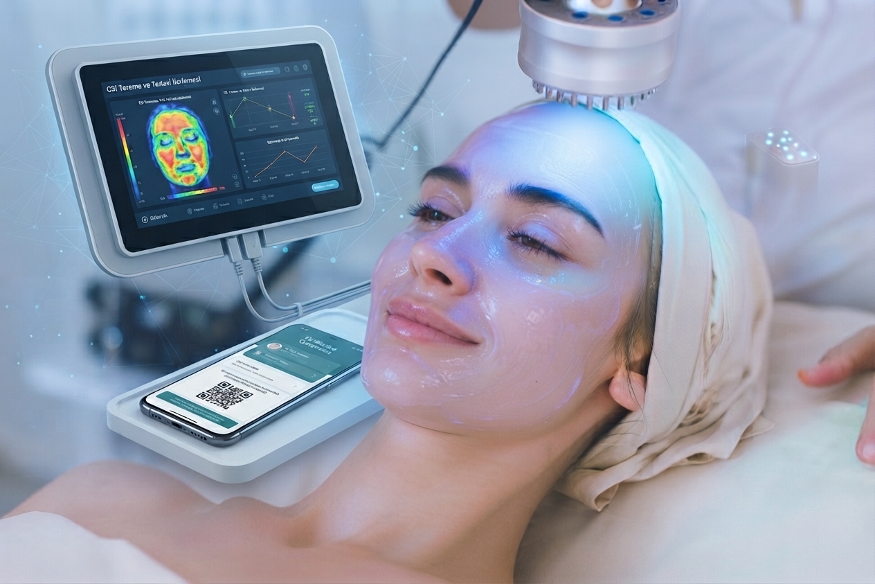
AI-Based Mobile Photodynamic Therapy Platform
Contactless imaging, PpIX fluorescence analysis and large language model (LLM) inference for lesion planimetry, treatment response, outcome prediction and personalized PDT protocol.
Developed for German university clinics.
PlanimetryPpIX FluorescenceTreatment ResponseOutcome PredictionTeleconsultation
Table of Contents
1What is AI PDT Analysis?
2Technology: Image Processing & LLM Inference
37 Modules & Evaluated Parameters
4Clinical Value & Decision Support
5ESG & Sustainability
6Scientific References
Vision
A software-centric, hardware-independent photodynamic therapy intelligence platform that produces clinically meaningful PDT assessment from consumer devices (smartphone, tablet).
Use Cases
University clinics, dermatology oncology centers, telemedicine and home care — 7-module AI-powered PDT assessment and tracking.
1
Section 1
What is AI PDT Analysis?
& Treatment Response Prediction Platform — PhotoGuard AI is an AI-powered mobile imaging support tool that non-invasively assesses lesion planimetry, PpIX fluorescence, treatment response, outcome prediction and personalized PDT protocol from a standard lesion photograph. The system analyzes lesion area, PpIX distribution, erythema, crusting and epithelialization.
Post-PDT tissue color and geometric changes alter light reflection/scattering and fluorescence characteristics. The AI quantifies these optical signatures through image processing and machine learning to produce lesion area (cm²), PpIX score, response score and clearance probability.
Core Capabilities
- Planimetry: lesion area, perimeter, depth — with reference card calibration
- PpIX Fluorescence: intensity score (0-100), distribution pattern, incubation compliance
- Treatment Response: erythema, crusting, necrosis (%), epithelialization (%)
- Outcome Prediction: predicted sessions, clearance probability (%), recurrence risk
- Personalized Protocol: photosensitizer, light dose, wavelength, session interval
- Allergen & Light Protection: allergen-safe photosensitizer, SPF 50+ plan
- Teleconsultation: anonymized submission, urgency triage
Clinical Positioning
The system is a screening and decision-support tool; it does not replace a definitive medical diagnosis. AI models may err; outputs are produced to support a dermatologist/oncologist's clinical assessment. Final diagnosis and treatment decisions rest with the specialist physician.
2
Section 2
Technology: Image Processing & LLM Inference
The platform combines image processing and large language model (LLM)-based multimodal inference. From a standard RGB image, the lesion region is segmented; tissue, color, fluorescence and geometric features are extracted and clinical inference is performed with a multimodal LLM.
Processing Flow
- Image capture & quality audit (brightness, contrast, sharpness, reference card detection)
- Lesion segmentation and planimetric measurement (area, perimeter, depth)
- PpIX fluorescence assessment: intensity score, distribution pattern, incubation compliance
- Multimodal LLM inference: treatment response, outcome prediction, personalized protocol
- Summary metrics: lesion_area_cm2, fluorescence_score, response_score, clearance_probability
Software-Centric Architecture
Hardware-Independent
Runs only on the device's built-in camera; no extra medical hardware required.
Web-based (SaaS)
API-first architecture; integrates with EMR and clinical record systems.
AI Integration
Image processing + multimodal LLM for lesion localization and tissue mapping.
DiGA-Ready
German digital health application (DiGA) compliance roadmap; MDR Class IIa target.
3
Section 3
7 Modules & Evaluated Parameters
The analysis provides multi-layer parameters across 7 specialized modules. Each parameter is reported as percentage (0-100), description, severity and risk level.
PDTTrack — Planimetry
Lesion area (cm²), perimeter (cm), depth (mm), session-based area reduction (%)
PDTFluorescence — PpIX
PpIX fluorescence score (0-100), distribution pattern, incubation compliance
PDTResponse — Response
Erythema, crusting, necrosis (%), epithelialization (%), response level
PDTPredict — Prediction
Predicted sessions, clearance probability (%), recurrence risk, stagnation warning
PDTProtocol — Protocol
Photosensitizer type, incubation time, light dose (J/cm²), wavelength (nm)
PDTSafety — Safety
Allergen-safe photosensitizer, light protection plan, photosensitivity risk score
PDTTeleConsult — Tele-Expert
Anonymized submission, urgency triage, specialist feedback
Output Format
- Track: lesion_area_cm2, lesion_perimeter_cm, lesion_depth_mm, area_reduction_pct
- Fluorescence: fluorescence_score, fluorescence_distribution, incubation_compliance
- Response: response_score, erythema_score, crusting_score, necrosis_pct, epithelialization_pct
- Predict: predicted_sessions, clearance_probability, recurrence_risk, stagnation_warning
- AI clinical commentary + personalized PDT protocol + printable full report (PDF)
4
Section 4
Clinical Value & Decision Support
The platform supports clinical workflows as a screening and pre-triage tool. Lesion parameter mapping provides structured, reproducible and comparable data to the physician. Final diagnosis and treatment decisions always rest with the specialist physician.
Screening
Clinical/home-care non-invasive PDT assessment support.
Decision Support
Structured response/outcome scores + clearance prediction + AI commentary.
Tracking
Longitudinal trend and lesion reduction curve monitoring.
Safety Architecture (Human-in-the-loop)
- AI output is not a diagnosis; it is information supporting the physician's assessment
- AI commentary does not contain prescription/treatment recommendations
- Per-patient data isolation (RLS) and KVKK/GDPR-aligned sensitive-data approach
Integration Scenarios
University Clinics
Longitudinal PDT tracking in dermatology/oncology clinics.
Telemedicine & Home Care
Remote consultation, recording from mobile devices and tracking.
5
Section 5
ESG & Sustainability
The software-centric architecture aligns with European Union carbon-reduction and ESG (Environmental, Social, Governance) goals. Hardware independence brings medical consumable use close to zero and reduces the carbon footprint from patients' physical travel through telemedicine.
Zero Hardware
Eliminates carbon from production-to-disposal of specialized PDT imaging devices.
Zero Consumables
No single-use probes/apparatus; clinical waste near zero.
Digital Logistics
Web-based analysis removes the need for patient travel to the clinic.
Roadmap
Phase 1
Clinical deployment of 7-module PDT assessment platform in university clinics.
Phase 2
Deep-learning-based automated lesion segmentation and response model.
Phase 3
DiGA approval and personalized PDT care suite.
6
Section 6
Scientific References
The following studies establish the clinical validity and methodological foundations of image-based AI assessment of photodynamic therapy and lesion evaluation. Our PhotoGuard AI platform draws inspiration from this literature but does not replace these methods.
1.Szeimies RM, et al. Photodynamic therapy for non-melanoma skin cancer. Lancet Oncol. 2002;3(5):289-295. DOI: 10.1016/S1470-2045(02)00737-6
2.Braathen LR, et al. Guidelines on the use of photodynamic therapy for nonmelanoma skin cancer. J Am Acad Dermatol. 2007;56(1):125-143. DOI: 10.1016/j.jaad.2006.06.006
3.Morton CA, et al. European guidelines for topical photodynamic therapy part 1. J Eur Acad Dermatol Venereol. 2019;33(1):14-29. DOI: 10.1111/jdv.15257
4.Morton CA, et al. European guidelines for topical photodynamic therapy part 2. J Eur Acad Dermatol Venereol. 2020;34(1):17-29. DOI: 10.1111/jdv.16081
5.Serra-Guillén C, et al. Update on photodynamic therapy for skin cancer. Actas Dermosifiliogr. 2019;110(5):361-369. DOI: 10.1016/j.ad.2018.10.001
6.Babilas P, et al. Photodynamic therapy in dermatology. Eur J Dermatol. 2010;20(6):723-734. DOI: 10.1684/ejd.2010.1082
7.Wiegell SR, et al. Changes in PpIX fluorescence during PDT. J Dermatol Sci. 2011;62(1):46-52. DOI: 10.1016/j.jdermsci.2011.01.003
8.Ericson MB, et al. Photodynamic therapy — fluorescence and clinical outcome. J Dermatol Sci. 2008;50(3):215-222. DOI: 10.1016/j.jdermsci.2008.01.011
9.Fritsch C, et al. Photodynamic diagnosis and therapy in dermatology. Skin Pharmacol Appl Skin Physiol. 2001;14(6):351-362. DOI: 10.1159/000056418
10.Lange C, et al. Machine learning for skin lesion analysis. J Eur Acad Dermatol Venereol. 2020;34(7):1435-1443. DOI: 10.1111/jdv.16415
11.Esteva A, et al. Dermatologist-level classification of skin cancer with deep neural networks. Nature. 2017;542(7639):115-118. DOI: 10.1038/nature21056
12.Haenssle HA, et al. Man against machine: diagnostic performance of a deep learning CNN. Ann Oncol. 2018;29(8):1837-1845. DOI: 10.1093/annonc/mdy166
13.Wang J, et al. Artificial intelligence in dermatology. J Dermatol. 2021;48(1):23-32. DOI: 10.1111/1346-8138.15616
14.Salim A, et al. Image-based AI assessment of skin lesions. Comput Biol Med. 2022;141:105115. DOI: 10.1016/j.compbiomed.2021.105115
PhotoGuard AI — Photodynamic Therapy & Lesion Assessment Platform
For German university clinics · Contactless imaging · AI decision support · odiosoft.tech